Also color-blind in mechanism, since the energy is delivered through insulated needles below the epidermis. The needle injury itself can provoke pigment change, so conservative density and good aftercare matter.
The family is widely used on darker skin.
Long-wavelength lasers
The 1,064 nanometer neodymium YAG laser sits far enough into the infrared that melanin absorbs it weakly. That is why it is the standard choice for hair removal and vascular treatment in types IV to VI.
The non-ablative fractional lasers at 1,550 nanometers are used across the range with reduced density and longer intervals. The 1,927 nanometer thulium laser is used with care, because its superficial absorption heats the epidermis despite the long wavelength.
Subdermal lasers
Devices such as Endolift deliver their wavelength beneath the epidermis through a fiber, so the melanin layer is bypassed. The entry points and the heated field can still pigment.
The Endolift guide covers that risk.
Ablative fractional lasers
Carbon dioxide and erbium resurfacing injure the epidermis by design, and in deeper skin that injury pigments with some regularity. Erbium, with its lower thermal spread, is the less risky of the two.
Many providers will not perform fractional CO2 above type IV. Those who do use low density, careful pretreatment and long follow-up, and the resurfacing guide covers the choice.
Intense pulsed light and short-wavelength lasers
At the top of the risk list. IPL's broad spectrum includes the wavelengths melanin absorbs most, and the device cannot distinguish the melanin in a spot from the melanin in the skin around it.
Short-wavelength lasers such as the 532 nanometer KTP and the 755 nanometer alexandrite have the same problem to a lesser degree. Type IV skin is treated by some providers with long cutoff filters, low fluence and a test spot.
Types V and VI are generally declined. A clinic that offers an IPL photofacial to a type VI face without hesitation has not understood its own device, and the IPL guide sets out the alternatives.
What a careful provider does differently
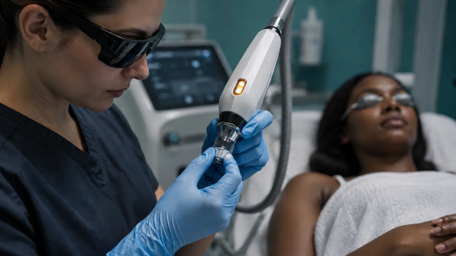
Cooling first. Contact cooling through a chilled sapphire tip, cryogen spray timed before the pulse, or forced cold air across the skin all pull heat out of the epidermis while the target heats.
A handpiece that feels warm rather than cold is a warning.
Longer pulse durations spread the energy over time. The small melanin granules in the epidermis cool between the pulse's start and finish while the larger target does not.
Lower fluence and lower fractional density reduce the total heat load, at the cost of more sessions. Longer intervals between sessions let pigment settle before the next insult.
The test spot is the single most useful practice. A small area behind the ear or under the jaw is treated at the proposed settings, and the provider looks at it one to four weeks later before treating the face.
It costs a visit. It has prevented a great many dark patches.
Pretreatment with a topical lightening agent for several weeks before resurfacing, under the direction of the treating physician, is common practice for deeper tones and reduces the pigment response.
Strict sun avoidance before and after treatment matters more for this group than for any other. The aftercare guide covers it day by day.
The questions to ask
How many people with my skin type have you treated on this specific device in the past year? What settings do you use for my type, and how do they differ from your settings for a type II?
Will you do a test spot, and how long will you wait before treating my face? What cooling does this device use?
What is your rate of pigment change in skin like mine, and what do you do when it happens? Can I see before-and-after photographs of people with my skin tone, taken under the same lighting?
The last question deserves a note. Photographs of darker skin under different lighting are nearly useless.
A clinic whose gallery is entirely pale faces is telling you something about its experience, whether it means to or not. Put the same questions to every practice under consideration and compare the answers side by side.
Some local context
Los Angeles has excellent public science institutions, and several of them explain this physics better than any clinic handout. The absorption of light by pigment is standard optics.
The public lectures and outreach material from Caltech in Pasadena cover why different wavelengths behave differently in different materials, with no treatment attached at the end.
Its neighbor up the hill works on the same physics at a different scale. Instruments that read the composition of a distant surface from the light it absorbs rest on the same absorption curves a laser operator should know.
The public pages of NASA's Jet Propulsion Laboratory explain that spectroscopy in plain language, and a reader who follows them will never again accept "it targets the pigment" as a full answer.
None of this is required reading before a consultation. It is useful reading, because a provider's explanation of wavelength and melanin is easier to judge once the physics is familiar.
Readers who learn better by watching than by reading have options too. Science programming on PBS has covered both skin pigmentation and laser physics with a care that aesthetic marketing rarely matches.
A recovery week is also a week to keep a healing face out of the sun, and a family day indoors is one way to spend it. Museums with a light and color gallery are the natural choice.
The light and color exhibits at the California Academy of Sciences are a long drive north, and a good reason to take one.
Bench summary
Melanin competes for light, so every light-based device carries more surface risk in deeper skin. The risk rises from the long-wavelength lasers through the ablative lasers to IPL at the top.
Radiofrequency, ultrasound and subdermal devices sidestep the problem. A careful provider cools, lengthens pulses, lowers fluence and density, spaces sessions, pretreats, and does a test spot.
Ask for the count, the settings, the photographs and the test spot. Treat a clinic's confidence as no substitute for any of them.